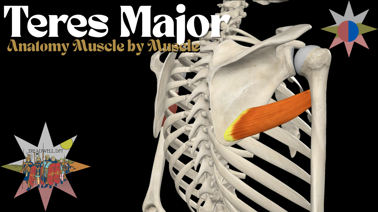
Teres Major Muscle – Anatomy Breakdown Video & Clinical Guide
The Teres Major is often called the "little lat" for its similar role in shoulder movement, but understanding its anatomy and function is crucial for effective assessment and rehab. In this video, we’ll break down its origin, insertion, function, innervation, and clinical relevance. Perfect for students, clinicians, trainers, and anyone wanting a clear, practical understanding of shoulder anatomy.
Watch the full video below and read on for more detail.
Quick Hits
The Teres Major is a thick, rounded muscle of the posterior shoulder that works closely with the latissimus dorsi to control internal rotation and adduction.
Origin: Posterior aspect of the inferior angle of the scapula.
Insertion: Medial lip of the intertubercular sulcus (bicipital groove) of the humerus.
Innervation: Lower subscapular nerve (C5, C6).
Actions: Shoulder internal rotation, adduction, and extension from a flexed position.
A major synergist to the latissimus dorsi, it assists in powerful pulling and climbing motions.
Clinical Relevance
Clinically, Teres Major tightness or overactivity can limit shoulder mobility, especially external rotation and overhead elevation.
It can contribute to altered scapulohumeral rhythm and compensatory movement patterns in athletes and lifters. Overdeveloped teres major relative to external rotators can predispose to shoulder impingement or internal rotation bias.
Rehabilitation often includes dry needling, soft tissue work, and balanced strengthening of the external rotators and scapular stabilizers. Proper movement cueing in pulling exercises is essential to avoid excessive internal rotation dominance.
What’s Next
𖤓Want help with shoulder pain?
I offer virtual consultations and personalized treatment plans for musculoskeletal issues!
𖤓In the Minneapolis area looking for treatment?
Book an appointment here!
𖤓Enjoyed the video?
There’s more where that came from! Check out my YouTube channel here!

