
Vastus Medialis – Anatomy Breakdown
Treadwell, DPT | Muscle by Muscle Series
Watch the Episode
Watch on YouTube: Vastus Medialis – Anatomy Breakdown
In this episode, Dr. Austin Treadwell, DPT breaks down the Vastus Medialis — the teardrop-shaped medial head of the quadriceps, best known for its role in patellar stability, knee extension, and that clean finish in terminal range.

Overview
The Vastus Medialis sits on the inner thigh, running from the femur to the patella, where its distal fibers (the Vastus Medialis Oblique, or VMO) insert obliquely to provide critical medial pull on the patella.
It’s smaller in volume than the Vastus Lateralis, but its function is just as vital — it balances lateral forces at the knee, keeping patellar motion smooth and centered during extension.
Clinically, it’s the go-to muscle for addressing patellofemoral pain, knee instability, and quad symmetry post-ACL injury.
Origin & Insertion
Origin: Medial lip of the linea aspera and intertrochanteric line of the femur
Insertion: Medial border of the patella → continues via the patellar ligament to the tibial tuberosity
The VMO fibers angle medially (≈50–55°), giving them leverage to counteract the lateral pull of the Vastus Lateralis and IT band.
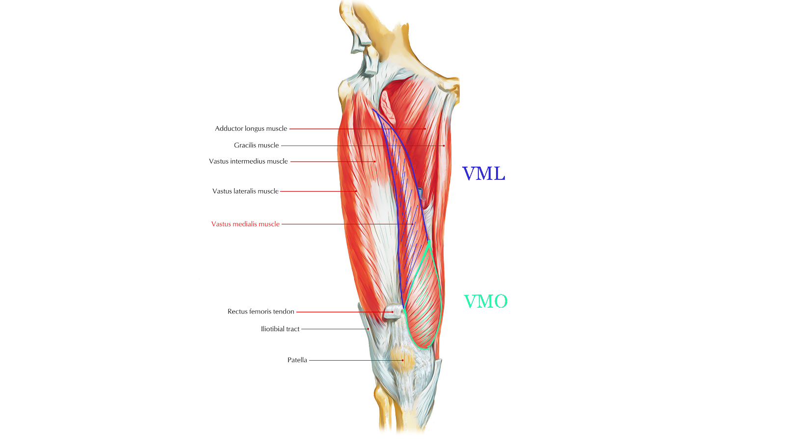
An anatomical diagram illustrating the Vastus Medialis Longus (VML) and Vastus Medialis Oblique (VMO) — the two segments of the Vastus Medialis muscle.
The VML fibers run more vertically, contributing to knee extension, while the VMO fibers run at a sharper medial angle (~50–55°), providing a medial stabilizing pull on the patella.
Together, these segments maintain balance against the lateral pull of the Vastus Lateralis and IT band, essential for smooth patellar tracking and controlled knee mechanics.
Function
Primary: Knee extension
Secondary: Medial stabilization of the patella and control of terminal extension (“last 15 degrees”)
Dynamic Role: Guides patellar tracking through the trochlear groove
Biomechanical data show that Vastus Medialis activation peaks between 0°–15° of knee flexion — the range where lateral translation risk is highest (Elias et al., 2022).
That’s why it’s the focus of most terminal knee extension (TKE) drills in rehab.
Innervation & Blood Supply
Innervation: Femoral nerve (L2–L4)
Blood Supply: Femoral and popliteal arteries
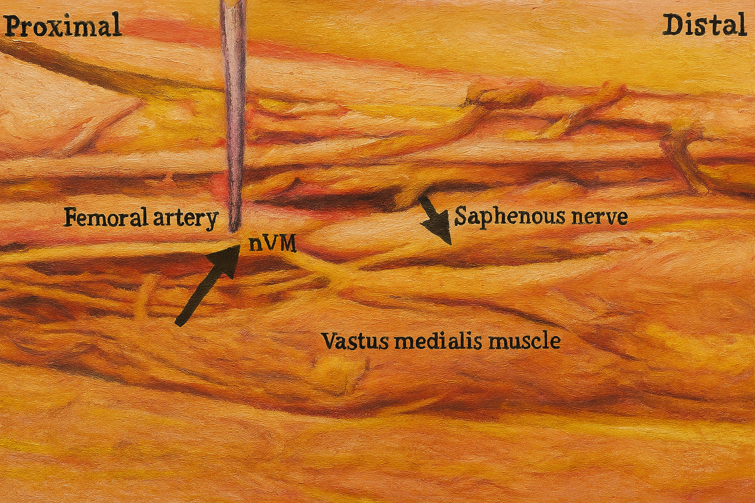
A labeled anatomical dissection highlighting the nerve to the Vastus Medialis (nVM) in proximity to the femoral artery and saphenous nerve within the distal thigh.
This visualization emphasizes the neurovascular anatomy of the medial thigh, critical for understanding VMO activation, surgical approaches, and nerve preservation during ACL or medial knee procedures.
Clinically, it underscores how nVM integrity influences medial patellar control and quadriceps reactivation following injury or surgery.
Clinical & Training Insights
The Vastus Medialis is a stability specialist — not just a strength muscle.
Weakness or delayed firing leads to poor patellar control, often presenting as “knee drift” or anterior knee pain.
Eccentric control during knee flexion is as important as concentric power during extension.
EMG studies confirm selective VMO engagement during terminal knee extension, mini-squats, and step-downs with external rotation (Cerny et al., 1995).
Volume Note: While the Vastus Medialis is smaller, it still plays a crucial role — making up roughly 25% of total quadriceps volume (Ema et al., 2017).
Clinical insight: Strength symmetry between the Vastus Medialis and Lateralis is a key predictor of patellar tracking health — imbalance in either direction increases PFJ stress and pain.

A labeled anatomical depiction highlighting the Vastus Medialis Oblique (VMO) in relation to the Medial Collateral Ligament (MCL), Medial Patellofemoral Ligament (MPFL), and Adductor Tendon. This region plays a vital role in patellar stability and medial knee control, helping counteract lateral tracking forces on the patella during extension.
This image is ideal for illustrating the anatomical integration of the VMO with medial knee structures relevant to patellofemoral mechanics and rehab.
Clinical Relevance
The Vastus Medialis is often the first to go and last to come back in knee rehab.
Its loss of tone post-injury can lead to compensations throughout the kinetic chain — from hip rotation to tibial alignment.
Early reactivation, controlled TKEs, and coordinated hip–quad co-contraction drills are essential for restoring functional stability.
Take the Next Step
You’ve got the anatomy down — now put it into motion.
If you’re a clinician, let’s talk patellofemoral rehab and quad sequencing.
If you’re an athlete or lifter, let’s talk knee tracking and end-range control.
And if you’re rebuilding after injury — this is where anatomy meets recovery.
𖤓 Watch more breakdowns on YouTube: Treadwell, DPT – Muscle by Muscle Series
𖤓 Book a Virtual Consultation: TreadwellDPT.com/appointments
𖤓 Download free PT tools & anatomy resources: TreadwellDPT.com/resources
Much more in store; even more to come.
Stay tuned, stay locked. Treadwell, DPT. 🏹