
The fibularis longus, also known as the peroneus longus, is a muscle located within the lateral compartment of the lower leg.
It begins along the outside of the leg, travels behind the lateral ankle, crosses underneath the foot, and inserts along the medial side of the first ray. This long, winding path allows the fibularis longus to influence the ankle, foot, and arch as one connected system.
The muscle contributes to ankle eversion and plantarflexion, but its role extends beyond producing movement. It helps stabilize the lateral ankle, support the foot’s arches, control the first ray, and create a strong lever during walking, running, jumping, and cutting.
Watch the Fibularis Longus Anatomy Breakdown: https://youtu.be/6sffn7uo8gU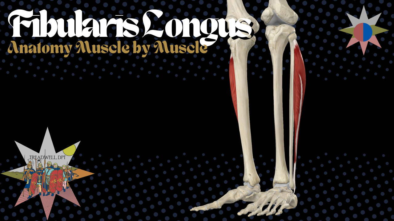
In this episode of Anatomy Breakdown – Muscle by Muscle, we cover:
Fibularis longus origin and insertion
Its path behind the lateral malleolus
Its route underneath the foot
Ankle eversion and plantarflexion
First-ray stabilization
Arch support and push-off mechanics
Innervation and nerve roots
Lateral ankle sprains
Chronic ankle instability
Dynamic ankle rehabilitation
What Is the Fibularis Longus?
The fibularis longus is one of the primary muscles within the lateral compartment of the lower leg.
The lateral compartment mainly contains:
Fibularis longus
Fibularis brevis
The fibularis longus is the more superficial of the two muscles and begins higher along the fibula. The fibularis brevis sits deeper and originates farther down the lower leg.
The names help distinguish them:
Longus means long.
Brevis means short.
The fibularis longus is therefore the longer fibularis muscle.
Fibularis Longus vs. Peroneus Longus
Fibularis longus and peroneus longus are two names for the same muscle.
The term peroneus comes from an older Greek-derived naming convention, while fibularis refers directly to the fibula.
Both names may still appear in anatomy resources, clinical documentation, and educational materials.
Modern anatomical terminology generally favors:
Fibularis longus
However, someone who learned the older terminology may know it as:
Peroneus longus
Fibularis Longus Origin
The fibularis longus originates from the:
Head of the fibula
Proximal portion of the lateral fibular shaft
This places the muscle high along the outside of the lower leg.
From its origin, the muscle belly travels down through the lateral compartment before narrowing into a long tendon.
Fibularis Longus Insertion
The fibularis longus inserts onto the:
Base of the first metatarsal
Medial cuneiform
Although the muscle begins on the lateral side of the lower leg, it ultimately inserts on the medial side of the foot.
Its tendon follows a distinctive path:
Travels down the lateral lower leg.
Passes behind the lateral malleolus.
Continues along the lateral side of the foot.
Runs through a groove associated with the cuboid.
Crosses underneath the foot.
Inserts onto the first metatarsal and medial cuneiform.
In simple terms:
Proximal fibula to first metatarsal and medial cuneiform
This creates lateral-compartment anatomy with significant medial-foot influence.
Fibularis Longus and the Lateral Malleolus
The fibularis longus tendon passes behind the:
Lateral malleolus
The fibularis brevis tendon also passes through this region.
The two tendons are held in place by the superior fibular retinaculum.
This retinaculum helps prevent the tendons from moving excessively around the back of the lateral ankle.
If the retinaculum is injured, the fibularis tendons may become unstable and can occasionally sublux or snap around the lateral malleolus.
Fibularis Longus Function
The primary actions of the fibularis longus include:
Ankle eversion
Ankle plantarflexion
Eversion turns the sole of the foot outward.
Plantarflexion points the foot downward at the ankle.
Because the fibularis longus tendon passes behind the ankle joint, it can assist with plantarflexion. However, its most recognized ankle function is eversion.
Fibularis Longus and Ankle Eversion
The fibularis longus helps pull the foot into eversion and resist excessive inversion.
This is important because a typical lateral ankle sprain occurs when the foot moves rapidly into inversion, often with some degree of plantarflexion.
The lateral ankle ligaments provide passive stability, including the:
Anterior talofibular ligament
Calcaneofibular ligament
Posterior talofibular ligament
The fibularis muscles provide active, contractile support.
They help respond when the foot begins rolling inward and contribute to the body’s ability to control ankle position during movement.
Fibularis Longus and the First Ray
The first ray generally refers to the functional unit involving the first metatarsal and medial cuneiform.
Because the fibularis longus inserts onto these structures, it helps:
Plantarflex the first ray
Stabilize the medial column
Control the big-toe side of the foot
Support force transfer during push-off
Prepare the foot to act as a rigid lever
This is particularly important during the later stages of walking and running.
As the body moves forward over the foot, the first ray must accept load and remain controlled so force can pass efficiently through the big toe.
Fibularis Longus and Arch Support
The fibularis longus contributes to support of the:
Transverse arch
Medial longitudinal arch
Medial column of the foot
Its tendon crosses from the lateral side of the foot toward the medial first ray.
This creates a sling-like mechanical effect underneath the foot.
The muscle works with other structures, including the tibialis posterior, intrinsic foot muscles, plantar fascia, and surrounding ligaments, to help control the shape and stiffness of the foot during weight-bearing.
The fibularis longus does not support the arch by itself, but it is an important part of the larger system.
Fibularis Longus and Push-Off
During walking and running, the foot must transition from a flexible structure that accepts load into a stronger lever that supports propulsion.
The fibularis longus contributes by:
Stabilizing the first ray
Supporting the medial column
Controlling foot position
Assisting plantarflexion
Helping create a stable base for big-toe push-off
This makes the muscle relevant during:
Walking
Running
Sprinting
Jumping
Cutting
Stair climbing
Direction changes
Athletic push-off
A poorly controlled first ray can reduce the efficiency of force transfer through the foot.
Fibularis Longus Innervation
The fibularis longus is innervated by the:
Superficial fibular nerve
This nerve is also called the:
Superficial peroneal nerve
The associated nerve roots are primarily:
L5 and S1
The superficial fibular nerve supplies both major muscles of the lateral compartment:
Fibularis longus
Fibularis brevis
It also provides sensation to much of the lower lateral leg and dorsum of the foot.
Superficial Fibular Nerve Symptoms
Weakness with ankle eversion does not automatically mean the fibularis longus itself is injured.
Possible contributors include:
Fibularis muscle weakness
Fibularis tendon pathology
Superficial fibular nerve irritation
Common fibular nerve involvement
L5 or S1 nerve-root involvement
Pain-related inhibition
Reduced motor control after an ankle sprain
Sensory symptoms along the lateral lower leg or top of the foot may increase suspicion of superficial fibular nerve involvement.
Symptoms and examination findings should be interpreted together rather than attributing all weakness to one muscle.
Fibularis Longus and Lateral Ankle Sprains
The fibularis longus is a major part of the ankle’s dynamic defense against inversion.
During a lateral ankle sprain, attention often centers on the ankle ligaments, especially the anterior talofibular ligament.
Ligament injury is important, but ankle stability also depends on:
Muscle strength
Reaction time
Balance
Proprioception
Motor coordination
Ability to control rapid loading
Confidence during movement
The fibularis longus and fibularis brevis help provide active support when the ankle begins moving into a potentially dangerous position.
What Is Chronic Ankle Instability?
Chronic ankle instability describes recurring episodes of the ankle giving way, repeated ankle sprains, or a persistent feeling of instability following an initial injury.
Possible features include:
Recurrent ankle sprains
Feeling that the ankle gives way
Reduced confidence on uneven surfaces
Impaired balance
Reduced eversion strength
Altered muscle activation
Difficulty with cutting or landing
Ongoing swelling or discomfort
Fear during athletic activity
Chronic ankle instability is not simply a stretched-ligament problem.
It may involve both mechanical and neurological changes.
Fibularis Longus and Chronic Ankle Instability
Research suggests that people with chronic ankle instability may demonstrate reduced eversion force compared with people without instability.
This means the muscles responsible for resisting inversion may have a true strength deficit.
Research has also identified altered regional activation within the fibularis longus during ankle eversion.
This suggests the problem may involve more than generalized weakness. The muscle may be recruited differently after repeated ankle sprains or ongoing instability.
Potential impairments may include:
Reduced strength
Delayed activation
Altered recruitment patterns
Impaired reflex responses
Reduced muscular endurance
Poor control during fatigue
Difficulty responding to sudden ankle movement
Fibularis Muscle Size and Ankle Stability
Muscle structure may also be related to perceived ankle stability.
Research has associated better self-reported ankle stability with a larger total cross-sectional area of the fibularis muscles.
This does not prove that increasing muscle size alone will eliminate ankle instability.
It does support the importance of developing strength and capacity within the lateral compartment rather than focusing exclusively on the injured ligaments.
Fibularis Longus Rehabilitation
Early rehabilitation may include basic exercises such as:
Isometric ankle eversion
Banded ankle eversion
Controlled plantarflexion
Seated foot-strengthening exercises
Double-leg balance
Supported single-leg stance
These exercises can help restore initial strength and movement tolerance.
However, later rehabilitation should prepare the ankle for real-world demands.
Training the Fibularis Longus as a Stabilizer
Most ankle sprains do not happen during slow, controlled resistance-band exercises.
They occur when:
The foot lands unexpectedly
The body continues moving over the ankle
The athlete cuts or changes direction
The ankle becomes fatigued
The surface is uneven
The foot contacts another player
The ankle must react quickly
Later-stage training may therefore include:
Single-leg balance
Unstable or changing surfaces
Perturbation training
Lateral stepping
Hopping
Landing drills
Deceleration training
Cutting
Direction changes
Reactive balance
Sport-specific fatigue exposure
The goal is to train the lateral ankle muscles to respond when movement becomes unpredictable.
Fibularis Longus Exercises
Exercises that may strengthen the fibularis longus and lateral ankle system include:
Banded ankle eversion
Isometric eversion holds
Calf raises with controlled foot position
Single-leg calf raises
Single-leg balance
Lateral step-downs
Lateral hops
Forward and diagonal hopping
Balance with external perturbations
Cutting drills
Uneven-surface walking
Sport-specific agility work
Exercise selection should reflect the person’s current symptoms, injury stage, balance, strength, and athletic demands.
Can You Isolate the Fibularis Longus?
The fibularis longus and fibularis brevis both contribute to ankle eversion and are difficult to isolate completely.
The fibularis longus may be emphasized through activities involving:
Eversion
Plantarflexion
First-ray stabilization
Weight-bearing push-off
Arch control
The goal of rehabilitation is rarely perfect isolation.
A more useful goal is restoring the entire lateral ankle and foot system so it can produce force, control movement, and react to unexpected loading.
Clinical Takeaway
The fibularis longus is much more than a basic ankle evertor.
It contributes to:
Ankle eversion
Ankle plantarflexion
Resistance to excessive inversion
Dynamic lateral ankle stability
First-ray plantarflexion
Medial-column control
Transverse arch support
Efficient push-off
Balance and single-leg control
Its path from the lateral lower leg to the medial first ray allows it to influence the ankle and foot as one connected system.
When evaluating lateral ankle sprains, recurring instability, balance deficits, or poor push-off mechanics, the fibularis longus deserves close attention.
The ligaments matter, but the active muscular system must also be strong, coordinated, and prepared to respond when movement becomes unpredictable.
Frequently Asked Questions
What does the fibularis longus do?
The fibularis longus everts and plantarflexes the ankle. It also plantarflexes and stabilizes the first ray, supports the foot’s arches, and contributes to dynamic ankle stability.
Where is the fibularis longus located?
It is located within the lateral compartment of the lower leg, along the outside of the fibula.
What is the origin of the fibularis longus?
It originates from the head of the fibula and the proximal lateral portion of the fibular shaft.
Where does the fibularis longus insert?
It inserts onto the base of the first metatarsal and the medial cuneiform.
What nerve innervates the fibularis longus?
It is innervated by the superficial fibular nerve, primarily from L5 and S1.
Is fibularis longus the same as peroneus longus?
Yes. Fibularis longus and peroneus longus are two names for the same muscle.
Does the fibularis longus pass behind the ankle?
Yes. Its tendon passes behind the lateral malleolus before traveling along the lateral foot and underneath the cuboid.
Does the fibularis longus support the arch?
Yes. It contributes to transverse and medial arch control through its path beneath the foot and attachment to the first metatarsal and medial cuneiform.
What is the first ray?
The first ray generally refers to the first metatarsal and medial cuneiform functional unit along the medial side of the foot.
How does the fibularis longus help prevent ankle sprains?
It actively everts the ankle and helps resist excessive inversion. It also contributes to rapid muscular stabilization during landing, cutting, and unexpected foot movement.
Is fibularis longus weakness related to chronic ankle instability?
Reduced eversion strength and altered fibularis longus activation have been identified in people with chronic ankle instability.
What exercises strengthen the fibularis longus?
Banded eversion, single-leg balance, calf raises, lateral hopping, perturbation training, cutting drills, and reactive agility exercises can train the fibularis longus as part of the larger ankle-stability system.
Can the fibularis longus tendon snap behind the ankle?
The fibularis tendons can sublux or snap around the lateral malleolus if the supporting retinaculum is damaged or the tendons become unstable.
Continue Learning Anatomy
Continue exploring the origins, insertions, innervation, actions, and clinical relevance of the lower-leg muscles through Anatomy Breakdown – Muscle by Muscle.
Fibularis Brevis Anatomy
Continue with the shorter, deeper muscle of the lateral compartment.
Subscribe to Treadwell, DPT


Fabric:100% cotton
Fabric Weight:10.3 oz/yd² (350 g/m²)
Fabric Thickness:Thick
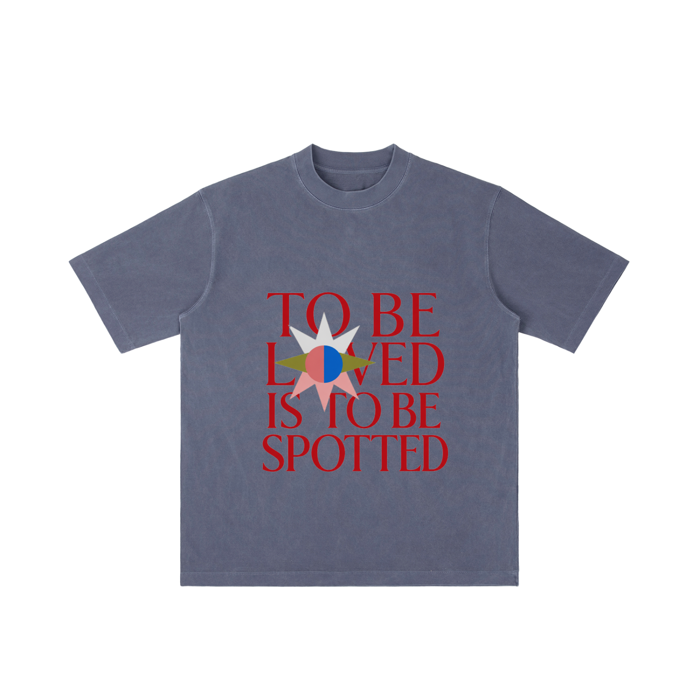
To be loved is to be spotted.
The To Be Loved Tee is a clean, minimal graphic shirt built around one of the realest gym truths: sometimes love looks like someone standing close, paying attention, and making sure you don’t get crushed under the bar.
With bold front typography and a simple eyes-and-hands back graphic, this tee blends gym culture, quiet romance, and modern streetwear energy into an easy everyday piece. It’s soft, simple, and just mysterious enough to make people look twice.
Perfect for lifters, gym couples, spotter-for-life relationships, fitness lovers, and anyone who knows being seen is part of being loved.
Best for:
Gym lovers, lifters, fitness couples, workout partners, streetwear fans, romantic graphic tee lovers, and anyone who appreciates a clever gym-coded message.
NO REFUNDS OR EXCHANGES
