
Video: https://youtu.be/jF5iW-KrjsE
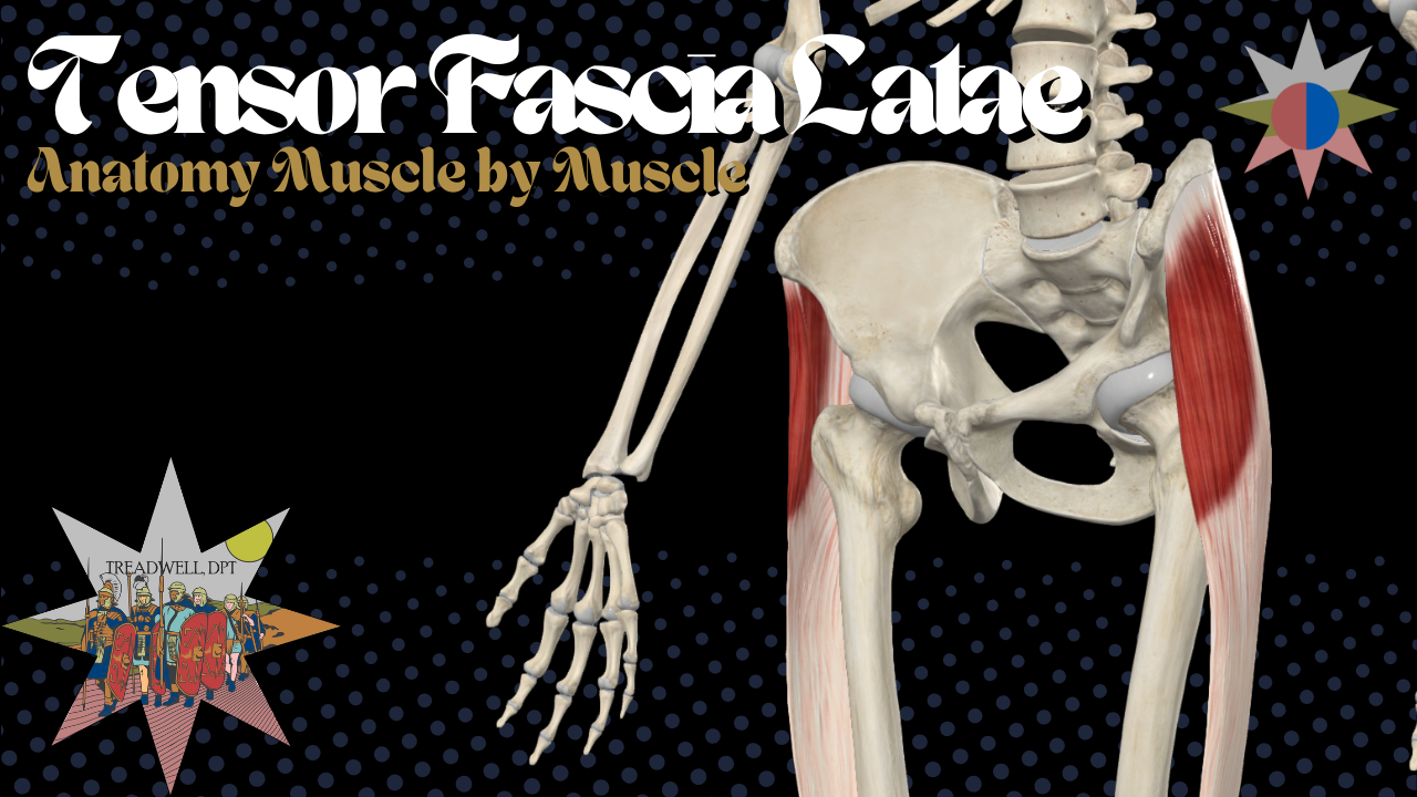
The tensor fasciae latae, commonly called the TFL, is a small muscle located along the front and outer portion of the hip. Despite its size, it plays an important role in hip movement, pelvic stability, walking, running, single-leg control, and tension through the iliotibial band.
In this Anatomy Breakdown, we cover the TFL’s:
Origin and insertion
Innervation
Primary actions
Role in hip and knee stability
Relationship with the iliotibial band
Clinical relevance
Stretching and self-management strategies
Watch the full video below for a clear, practical breakdown of tensor fasciae latae anatomy and function.
What Is the Tensor Fasciae Latae?
The tensor fasciae latae is a small, superficial muscle located on the anterolateral hip.
Its name helps describe its function:
Tensor means that it creates tension.
Fasciae latae refers to the deep fascia surrounding the thigh.
The TFL connects the pelvis to the iliotibial tract, commonly known as the IT band. Through this connection, the muscle can influence mechanics throughout the lateral hip, thigh, and knee.
Although the TFL is much smaller than muscles such as the gluteus maximus or gluteus medius, it contributes to several important lower-extremity movements and stabilization strategies.
Tensor Fasciae Latae Origin
The tensor fasciae latae originates from the:
Anterior superior iliac spine, or ASIS
Anterior portion of the iliac crest
These attachment points place the TFL along the front and outer portion of the pelvis.
Tensor Fasciae Latae Insertion
The TFL inserts into the:
Iliotibial tract
Also called the iliotibial band or IT band
The iliotibial tract continues down the lateral thigh and attaches to the lateral tibia at Gerdy’s tubercle.
Unlike muscles that insert directly onto one isolated bony landmark, the TFL blends into a large fascial structure. This allows it to influence tension and force transmission throughout the lateral thigh.
Tensor Fasciae Latae Innervation
The tensor fasciae latae is innervated by the:
Superior gluteal nerve
The associated nerve roots are generally:
L4, L5, and S1
The superior gluteal nerve also innervates the gluteus medius and gluteus minimus. These muscles work together during hip abduction, pelvic stabilization, walking, running, and single-leg activities.
Tensor Fasciae Latae Actions
The primary actions of the tensor fasciae latae include:
Hip flexion
Hip abduction
Hip internal rotation
The TFL helps move the thigh forward, move the leg away from the body, and rotate the femur inward.
Through its connection with the iliotibial band, it also contributes to lateral stabilization of the hip and knee.
Role of the TFL During Walking and Running
The tensor fasciae latae is not only responsible for producing movement. It also helps control movement.
The TFL can contribute during activities such as:
Walking
Running
Single-leg stance
Stair climbing
Landing
Cutting
Changing direction
Maintaining pelvic control
During single-leg tasks, the body must control the pelvis and femur over the planted leg. The TFL works alongside the gluteus medius, gluteus minimus, gluteus maximus, and other lower-extremity muscles to help manage these demands.
The TFL and the IT Band
The TFL inserts directly into the iliotibial band, making it an important part of the lateral hip and thigh system.
People frequently describe their IT band as being “tight.” However, the iliotibial band is a thick fascial structure rather than a muscle that can be stretched in the same way as contractile tissue.
Current biomechanical models increasingly describe the iliotibial band as a tensioned structure that can contribute to compression of underlying tissues. This is different from the traditional explanation that the IT band simply slides or rubs back and forth across the lateral femoral epicondyle.
Because the TFL adds tension to the iliotibial band, excessive TFL activity or poor load distribution may contribute to symptoms around the:
Anterolateral hip
Lateral thigh
Outer knee
Iliotibial band region
This does not necessarily mean that the TFL is damaged. It may mean that the muscle is performing more work than it should because other muscles or movement strategies are not contributing effectively.
Can the TFL Compensate for Weak Glutes?
The TFL can become dominant during exercises that are intended to strengthen the gluteal muscles.
For example, someone may perform hip abduction or single-leg exercises but rely heavily on the TFL rather than effectively recruiting the gluteus medius or gluteus maximus.
A TFL-dominant strategy may be associated with:
Tension near the front or outside of the hip
Excessive hip flexion
Excessive femoral internal rotation
Poor pelvic control
Difficulty controlling the knee during single-leg tasks
Recurrent lateral hip or thigh discomfort
The goal is not to completely shut off or eliminate TFL activity. The TFL is a normal and useful muscle.
Instead, rehabilitation and training should focus on improving how the entire system shares load.
That may include:
Improving gluteus maximus contribution
Improving gluteus medius strength and control
Training posterior-chain strength
Improving single-leg stability
Improving femoral and pelvic control
Modifying exercises that consistently produce unwanted TFL dominance
How to Stretch the Tensor Fasciae Latae
To place the TFL in a lengthened position, combine movements opposite its primary actions.
A TFL stretch may include:
Hip extension
Hip adduction
Slight hip external rotation
One simple standing TFL stretch is performed by:
Crossing the involved leg behind the other leg.
Shifting the hips toward the involved side.
Leaning the torso away from the involved side.
Maintaining a controlled position without twisting aggressively.
Holding the stretch while breathing normally.
The stretch should generally be felt near the front and outside of the hip.
Avoid forcing the movement or pushing into sharp pain.
Foam Rolling the TFL
Foam rolling may temporarily reduce discomfort or tension around the lateral hip, but aggressively rolling the full length of the IT band is not always necessary.
A more targeted approach is to focus near the TFL muscle belly, which is located close to the front and outer portion of the hip.
General foam-rolling guidelines include:
Use slow, controlled pressure.
Avoid aggressively rolling through significant pain.
Breathe normally and allow the area to relax.
Spend approximately 30 to 60 seconds on sensitive regions.
Stop if the technique produces sharp pain, numbness, or worsening symptoms.
Manual work and stretching may help manage symptoms, but they do not automatically correct the reason the TFL became overworked.
Long-term improvement usually requires better strength, movement control, exercise selection, and load management.
Clinical Takeaway
The tensor fasciae latae is a small but influential muscle.
It contributes to:
Hip flexion
Hip abduction
Hip internal rotation
Pelvic stability
Single-leg control
Lateral hip and knee stabilization
Tension through the iliotibial band
When someone complains of a “tight IT band,” the TFL and surrounding hip musculature may deserve closer attention.
Rather than trying to eliminate TFL activity, the goal should be to improve load distribution throughout the hip and lower extremity.
That means building strength where it is needed, improving pelvic and femoral control, and choosing exercises that encourage the desired muscular strategy.
Frequently Asked Questions
What does the tensor fasciae latae do?
The tensor fasciae latae assists with hip flexion, hip abduction, and hip internal rotation. It also contributes to pelvic stability and lateral stabilization of the hip and knee through its connection with the iliotibial band.
Where is the TFL located?
The TFL is located on the front and outer portion of the hip, near the anterior superior iliac spine and anterior iliac crest.
What nerve innervates the tensor fasciae latae?
The tensor fasciae latae is innervated by the superior gluteal nerve, primarily from the L4, L5, and S1 nerve roots.
Does the TFL attach to the IT band?
Yes. The TFL inserts into the iliotibial tract, commonly called the IT band.
Can a tight TFL cause IT band pain?
An overworked or sensitive TFL may increase tension through the iliotibial band and contribute to symptoms around the lateral hip, thigh, or knee. However, symptoms should be evaluated in the context of training load, hip strength, movement mechanics, and other possible sources of pain.
Can you stretch the IT band?
The IT band itself is a dense fascial structure and does not stretch like a muscle. Stretching positions may instead affect the TFL, gluteal muscles, and surrounding tissues.
How do you stretch the TFL?
A TFL stretch usually combines hip extension, hip adduction, and slight external rotation. One option is to cross the involved leg behind the other leg, shift the hips sideways, and lean the torso away from the involved side.
Should you foam roll the IT band?
Foam rolling may provide temporary symptom relief, but aggressive rolling directly along the entire IT band may be uncomfortable and unnecessary. Targeting the TFL and surrounding muscles may be more useful.
Continue Learning Anatomy
Explore more episodes of Anatomy Breakdown – Muscle by Muscle to learn the origins, insertions, innervation, actions, and clinical relevance of the muscles throughout the human body.
Subscribe on YouTube:
https://www.youtube.com/@TreadwellDPT
More physical therapy, anatomy, rehabilitation, and movement content:
https://TreadwellDPT.com
Fabric:100% cotton
Fabric Weight: 10.3 oz/yd² (350 g/m²)
Fabric Thickness: Thick
Physical therapy isn’t for the weak.
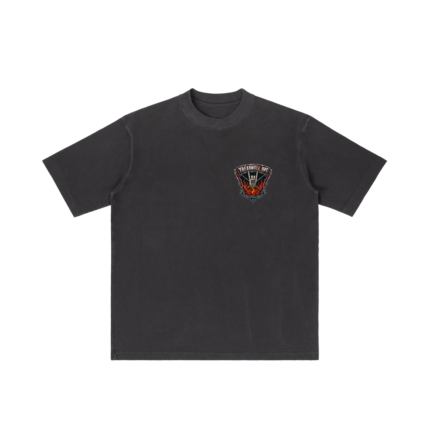
The I Survived Physical Therapy Tee is for anyone who’s been through the grind — the reps, the soreness, the “one more set,” and the moments where rehab felt like a full-contact sport. Built with a bold championship-style back graphic and a clean front badge, this shirt gives PT patients, rehab pros, and gym people a way to wear the comeback story.
Perfect for physical therapists, PT students, rehab clinics, post-op warriors, athletes, lifters, and anyone who earned the right to say they survived physical therapy.
Wear it after a hard session, gift it to a discharged patient, or rep it as a badge of honor for making it through the work.
Best for:
Physical therapy patients, PTs, PTAs, rehab pros, post-op recovery, athletes, gym lovers, and anyone who survived the treatment table.
NO REFUNDS/EXCHANGES
