
The fibularis tertius, also known as the peroneus tertius, is a small and highly variable muscle located within the anterior compartment of the lower leg.
Although it shares the fibularis name with the fibularis longus and fibularis brevis, it differs from both muscles in several important ways. It sits in a different compartment, receives a different nerve supply, crosses the front of the ankle, and assists with dorsiflexion rather than plantarflexion.
The fibularis tertius may also be absent in some people or blend closely with the extensor digitorum longus, making it one of the more anatomically interesting muscles of the lower leg.
Watch the Fibularis Tertius Anatomy Breakdown: https://youtu.be/5YeJc2bX_Ao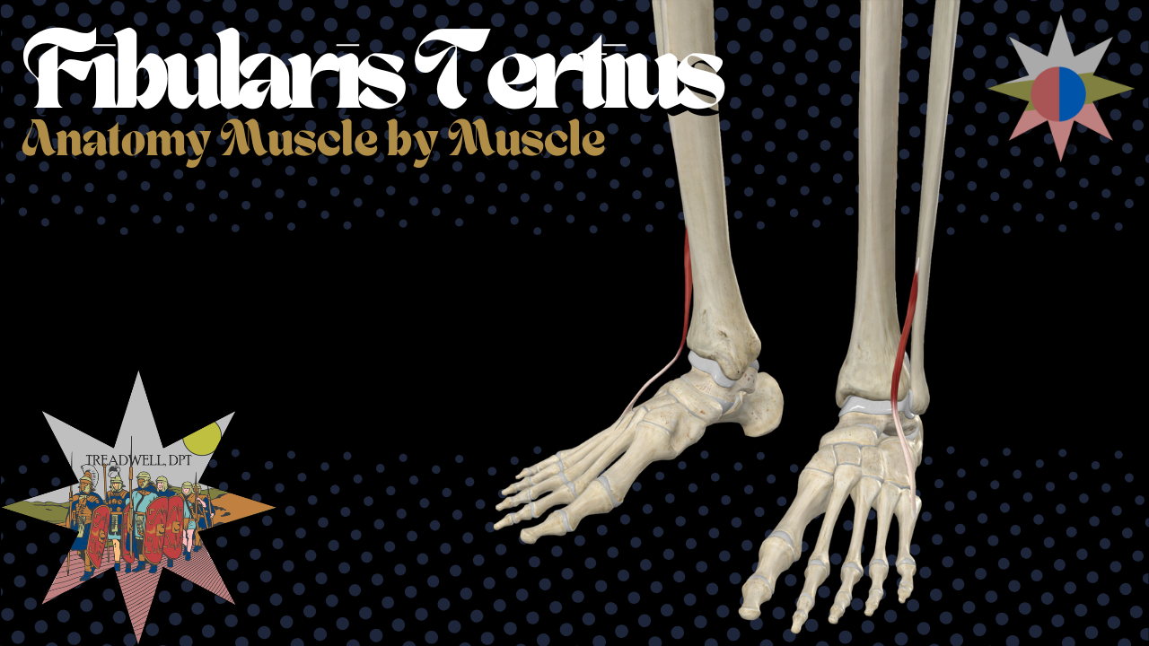
In this episode of Anatomy Breakdown – Muscle by Muscle, we cover:
Fibularis tertius origin and insertion
Its relationship with extensor digitorum longus
Its path across the front of the ankle
Ankle dorsiflexion and eversion
Innervation and nerve roots
Differences from fibularis longus and brevis
Anatomical variation and prevalence
Its possible role in foot clearance and walking
Developmental connections with extensor digitorum longus
What Is the Fibularis Tertius?
The fibularis tertius is considered the third fibularis muscle.
The name tertius means third:
Fibularis longus
Fibularis brevis
Fibularis tertius
However, the fibularis tertius does not sit in the same compartment as the other two muscles.
Fibularis longus and brevis are located within the lateral compartment of the lower leg. Fibularis tertius is located within the anterior compartment alongside the:
Tibialis anterior
Extensor hallucis longus
Extensor digitorum longus
This compartment difference helps explain its actions and nerve supply.
Fibularis Tertius vs. Peroneus Tertius
Fibularis tertius and peroneus tertius are two names for the same muscle.
The term peroneus comes from an older anatomical naming convention, while fibularis refers directly to the fibula.
Both names may still appear in:
Anatomy textbooks
Medical records
Research studies
Imaging reports
Clinical discussions
Modern anatomical terminology generally favors fibularis tertius.
Fibularis Tertius and Extensor Digitorum Longus
The fibularis tertius is closely related to the extensor digitorum longus.
It is often described as a partially separated portion of the extensor digitorum longus, almost as though the most lateral portion of that muscle developed into its own distinct structure.
This relationship helps explain why the fibularis tertius:
Sits within the anterior compartment
Shares the deep fibular nerve
Passes beneath the extensor retinaculum
Assists with dorsiflexion
May blend with extensor digitorum longus
Varies considerably between individuals
Although its name connects it with the other fibularis muscles, its anatomy and development may be more closely tied to the extensor digitorum longus.
Fibularis Tertius Origin
The fibularis tertius originates from the:
Distal third of the anterior fibula
Adjacent portion of the interosseous membrane
The interosseous membrane is a strong sheet of connective tissue extending between the tibia and fibula.
From its origin, the muscle runs downward through the anterior compartment before becoming a tendon near the ankle.
Fibularis Tertius Insertion
The fibularis tertius inserts onto the:
Dorsal surface of the base of the fifth metatarsal
The dorsal surface is the top of the foot.
Its tendon follows a short and relatively direct path:
Begins along the distal anterior fibula.
Travels through the anterior compartment.
Passes beneath the extensor retinaculum.
Crosses in front of the ankle joint.
Travels toward the outer portion of the foot.
Inserts onto the dorsal base of the fifth metatarsal.
In simple terms:
Distal anterior fibula to the dorsal base of the fifth metatarsal
Fibularis Tertius vs. Fibularis Brevis Insertion
The fibularis tertius and fibularis brevis both attach near the base of the fifth metatarsal, but they insert onto different surfaces and approach from different directions.
Fibularis Brevis
Passes behind the lateral malleolus
Approaches from the posterolateral ankle
Inserts onto the tuberosity at the base of the fifth metatarsal
Attaches more laterally and plantarly
Fibularis Tertius
Passes in front of the ankle
Approaches from the anterior compartment
Inserts onto the dorsal base of the fifth metatarsal
Attaches more superiorly on the foot
The two muscles therefore influence the same general region through different lines of pull.
Fibularis Tertius and the Extensor Retinaculum
The fibularis tertius tendon passes underneath the:
Extensor retinaculum
The extensor retinacula help secure the anterior-compartment tendons as they cross the front of the ankle.
This separates the fibularis tertius from the fibularis longus and brevis, whose tendons pass behind the lateral malleolus beneath the fibular retinacula.
The fibularis tertius remains anterior as it crosses the ankle joint.
That anterior path is the reason it contributes to dorsiflexion.
Fibularis Tertius Function
The primary actions of the fibularis tertius are:
Ankle dorsiflexion
Foot eversion
Dorsiflexion brings the top of the foot toward the shin.
Eversion lifts and turns the outer border of the foot upward.
The fibularis tertius contributes to dorsiflexion because its tendon passes in front of the ankle joint. It contributes to eversion because it travels laterally toward the fifth metatarsal.
Fibularis Tertius vs. Fibularis Longus and Brevis
All three fibularis muscles assist with eversion, but their paths around the ankle produce different secondary actions.
Fibularis Longus and Brevis
Pass behind the ankle joint
Assist with plantarflexion
Contribute substantially to eversion
Are innervated by the superficial fibular nerve
Fibularis Tertius
Passes in front of the ankle joint
Assists with dorsiflexion
Provides a smaller contribution to eversion
Is innervated by the deep fibular nerve
A useful comparison is:
Fibularis longus and brevis: eversion plus plantarflexion
Fibularis tertius: eversion plus dorsiflexion
The path around the ankle determines the action.
Fibularis Tertius and Tibialis Anterior
The fibularis tertius may help balance the inversion pull of the tibialis anterior during dorsiflexion.
The tibialis anterior is a strong dorsiflexor, but it also inverts the foot.
The fibularis tertius assists with dorsiflexion while pulling the foot toward eversion.
During ankle dorsiflexion:
Tibialis anterior pulls medially toward inversion.
Fibularis tertius pulls laterally toward eversion.
Together, they can help lift the foot while limiting excessive movement toward either side.
The fibularis tertius is not powerful enough to completely balance the tibialis anterior by itself, but it may provide a small fine-tuning contribution.
Is Fibularis Tertius a Strong Muscle?
The fibularis tertius is generally considered a relatively weak:
Dorsiflexor
Evertor
The tibialis anterior produces substantially more dorsiflexion force.
The fibularis longus and especially the fibularis brevis provide more significant eversion force.
The fibularis tertius is therefore best viewed as:
An assistant
A fine-tuning muscle
A contributor to lateral foot positioning
A small part of the anterior-compartment dorsiflexion system
It is not the primary muscle for either dorsiflexion or eversion.
Fibularis Tertius Innervation
The fibularis tertius is innervated by the:
Deep fibular nerve
This nerve is also called the:
Deep peroneal nerve
The associated nerve roots are primarily:
L5 and S1
The deep fibular nerve also supplies the other major muscles of the anterior compartment:
Tibialis anterior
Extensor hallucis longus
Extensor digitorum longus
Fibularis tertius
Deep Fibular Nerve Injury
Injury to the deep fibular nerve may contribute to weakness involving:
Ankle dorsiflexion
Great-toe extension
Lesser-toe extension
The fibularis tertius contribution to eversion
Depending on the level and severity of the injury, deep fibular nerve dysfunction may contribute to a foot-drop presentation.
Foot drop refers to difficulty lifting the front of the foot during walking.
The fibularis tertius is not the primary dorsiflexor, but weakness within the muscle may be one part of a larger anterior-compartment deficit.
Fibularis Tertius and Ankle Eversion Weakness
Most ankle eversion strength comes from the fibularis longus and fibularis brevis.
These muscles are supplied by the superficial fibular nerve.
However, the fibularis tertius demonstrates that not every ankle evertor shares that nerve supply.
When evaluating eversion weakness, possible contributors may include:
Fibularis longus weakness
Fibularis brevis weakness
Fibularis tertius weakness
Superficial fibular nerve dysfunction
Deep fibular nerve dysfunction
Common fibular nerve injury
L5 or S1 nerve-root involvement
Pain-related inhibition
The specific pattern of weakness and sensory change can help identify where the problem may be occurring.
Is Fibularis Tertius Present in Everyone?
The fibularis tertius is not present as a clearly distinct muscle in every person.
A large meta-analysis examining more than 7,600 legs found the muscle in approximately:
93% of adult cadaveric specimens
80% of clinical examinations
This difference may occur because the muscle can be:
Small
Difficult to palpate
Closely blended with extensor digitorum longus
Represented by a very thin tendon
Present anatomically but difficult to identify through the skin
Not seeing or feeling the tendon does not automatically mean the muscle is absent.
How to Find the Fibularis Tertius Tendon
To attempt to identify the fibularis tertius tendon:
Bring the foot upward toward the shin.
Pull the outer border of the foot slightly upward.
Combine dorsiflexion with eversion.
Look along the top and outer portion of the ankle.
Follow the tendon toward the dorsal base of the fifth metatarsal.
When visible, the tendon may appear along the dorsolateral ankle and foot.
However, it may remain difficult to distinguish from the nearby extensor digitorum longus tendons.
Fibularis Tertius Anatomical Variations
The fibularis tertius demonstrates considerable variation between individuals.
Its tendon may:
Insert onto the fifth metatarsal
Extend toward the fourth metatarsal
Attach to both the fourth and fifth metatarsals
Divide into multiple tendon slips
Form broader, fan-shaped attachments
Blend with nearby tendons or fascia
This means the muscle does not always appear as one small tendon inserting onto one precise point.
Its influence may extend across a wider portion of the dorsolateral foot.
Split Fibularis Tertius Tendon
A case report described a fibularis tertius tendon that divided into two separate slips.
One tendon slip continued toward the fifth toe.
The second connected with a plantar muscle along the lateral side of the foot.
The authors proposed that these additional attachments might contribute to:
Lateral arch support
Midfoot stabilization
Control of the lateral foot during walking
This type of variation is uncommon but demonstrates how variable the muscle can be.
Fibularis Tertius and Walking
The fibularis tertius may assist with foot clearance during the swing phase of walking.
During swing, the foot must lift away from the ground so the toes do not drag.
By assisting with dorsiflexion and lifting the outer border of the foot, the fibularis tertius may contribute to:
Foot clearance
Lateral-foot positioning
Preparing the foot for initial contact
Controlling excessive inversion during swing
Fine-tuning ankle position during gait
The muscle is not essential for normal walking. People without a distinct fibularis tertius can still walk normally.
Its role is more likely supportive than essential.
Is Fibularis Tertius Unique to Humans?
The fibularis tertius has historically been described as an exclusively human muscle.
More recent comparative anatomical research has made that claim less absolute.
However, the muscle continues to be discussed in relation to:
Upright posture
Bipedal locomotion
Foot clearance
Lateral-foot control
Human gait development
The ability to dorsiflex and evert the foot may provide a small advantage when positioning the foot during upright walking.
The fibularis tertius should therefore be viewed as a possible anatomical specialization rather than a requirement for bipedal gait.
Development of the Fibularis Tertius
Recent cadaveric research has further supported the close developmental relationship between the fibularis tertius and extensor digitorum longus.
The fibularis tertius was commonly supplied by a nerve branch shared with the extensor digitorum longus.
Researchers described the two as brother muscles, suggesting that they likely develop from the same original muscle mass.
The study also identified an inverse relationship between the size of the two muscles:
When fibularis tertius was larger, extensor digitorum longus tended to be smaller.
When fibularis tertius was absent, extensor digitorum longus extended farther down the leg.
This supports the idea that the fibularis tertius may represent a separated portion of the extensor digitorum longus.
Fibularis Tertius Exercises
Because the fibularis tertius assists with both dorsiflexion and eversion, exercises may combine the two movements.
Options may include:
Resisted ankle dorsiflexion
Resisted dorsiflexion with slight eversion
Toe raises
Heel walking
Controlled foot-clearance drills
Single-leg balance
Marching with active dorsiflexion
Gait drills
Step-over exercises
Anterior-compartment strengthening
Complete isolation is difficult because the tibialis anterior and extensor muscles will also contribute.
The goal is usually to strengthen and coordinate the anterior compartment rather than isolate the fibularis tertius perfectly.
Clinical Takeaway
The fibularis tertius is a small but anatomically distinctive muscle.
It contributes to:
Ankle dorsiflexion
Foot eversion
Lateral-foot positioning
Foot clearance during gait
Balance between inversion and eversion during dorsiflexion
Fine control of the ankle and foot
Unlike fibularis longus and brevis, it:
Sits in the anterior compartment
Crosses in front of the ankle
Assists with dorsiflexion
Receives the deep fibular nerve
Shares a close developmental relationship with extensor digitorum longus
It is present in most people, but its size, insertion, and visibility vary considerably.
The fibularis tertius is not the strongest dorsiflexor or evertor, but it provides a unique combination of both actions and helps fine-tune foot position during movement.
Frequently Asked Questions
What does the fibularis tertius do?
The fibularis tertius assists with ankle dorsiflexion and foot eversion. It may also help position the outer border of the foot during walking.
Where is the fibularis tertius located?
It is located within the anterior compartment of the lower leg alongside the tibialis anterior and long toe extensors.
What is the origin of the fibularis tertius?
It originates from the distal third of the anterior fibula and the adjacent interosseous membrane.
Where does the fibularis tertius insert?
It inserts onto the dorsal surface of the base of the fifth metatarsal.
What nerve innervates the fibularis tertius?
It is innervated by the deep fibular nerve, primarily from L5 and S1.
Is fibularis tertius the same as peroneus tertius?
Yes. Fibularis tertius and peroneus tertius are two names for the same muscle.
Is the fibularis tertius part of the lateral compartment?
No. Despite its name, it is located within the anterior compartment of the lower leg.
Does fibularis tertius plantarflex or dorsiflex the ankle?
It assists with dorsiflexion because its tendon crosses in front of the ankle joint.
How is fibularis tertius different from longus and brevis?
Fibularis longus and brevis pass behind the ankle, assist with plantarflexion, and receive the superficial fibular nerve. Fibularis tertius passes in front, assists with dorsiflexion, and receives the deep fibular nerve.
Is fibularis tertius present in everyone?
No. It is present in most people, but it may be absent, very small, or closely blended with extensor digitorum longus.
How do you test the fibularis tertius?
Ask the person to dorsiflex and evert the foot. The tendon may become visible along the dorsolateral ankle as it travels toward the fifth metatarsal.
Is fibularis tertius important for walking?
It may assist with foot clearance and lateral-foot positioning during the swing phase, but it is not essential for normal walking.
Can you isolate the fibularis tertius?
Complete isolation is difficult because other anterior-compartment muscles also assist with dorsiflexion.
Continue Learning Anatomy
Continue exploring the origins, insertions, innervation, actions, and clinical relevance of the lower-leg muscles through Anatomy Breakdown – Muscle by Muscle.
Fibularis Longus Anatomy
Fibularis Brevis Anatomy
Subscribe to Treadwell, DPT


Fabric:100% cotton
Fabric Weight:10.3 oz/yd² (350 g/m²)
Fabric Thickness:Thick
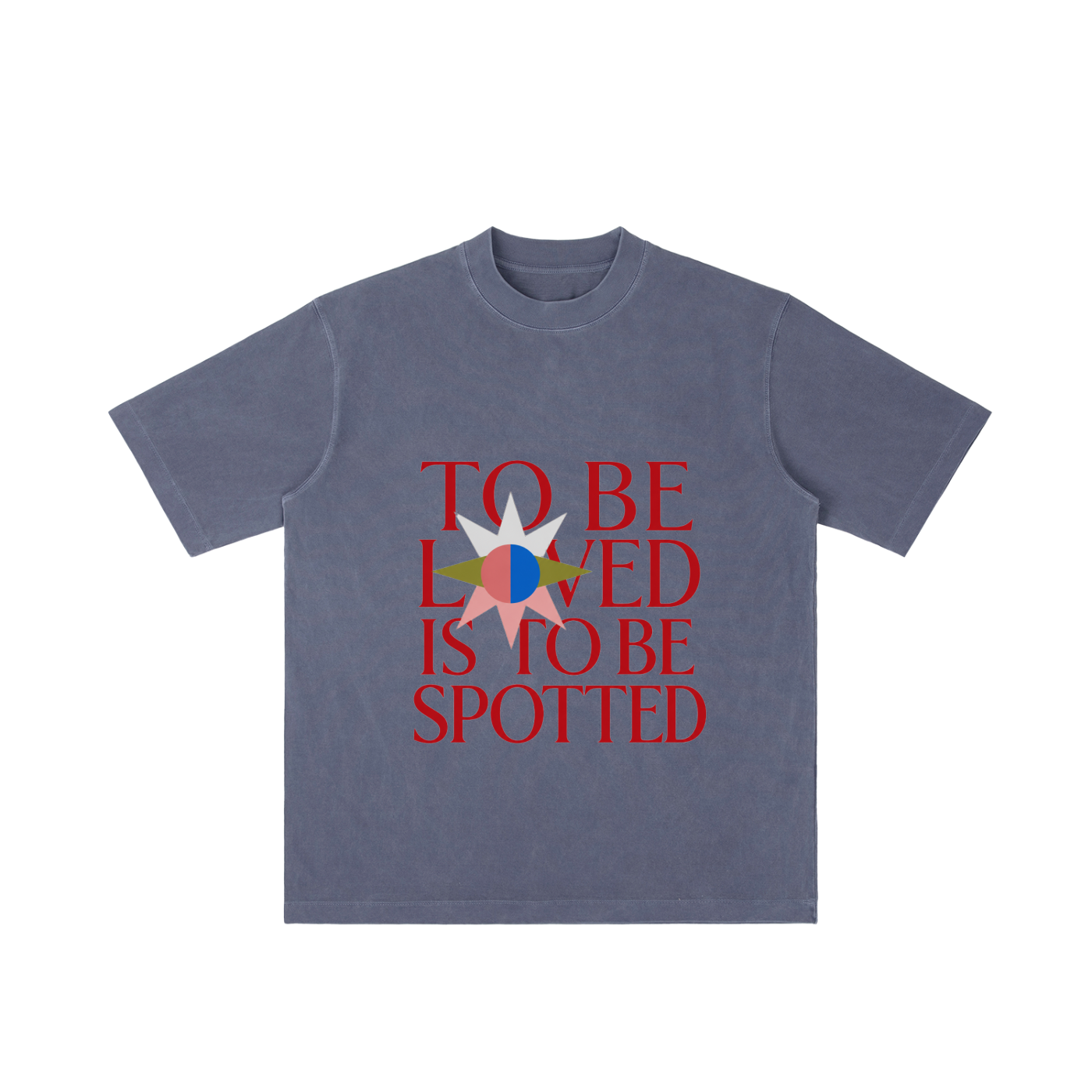
To be loved is to be spotted.
The To Be Loved Tee is a clean, minimal graphic shirt built around one of the realest gym truths: sometimes love looks like someone standing close, paying attention, and making sure you don’t get crushed under the bar.
With bold front typography and a simple eyes-and-hands back graphic, this tee blends gym culture, quiet romance, and modern streetwear energy into an easy everyday piece. It’s soft, simple, and just mysterious enough to make people look twice.
Perfect for lifters, gym couples, spotter-for-life relationships, fitness lovers, and anyone who knows being seen is part of being loved.
Best for:
Gym lovers, lifters, fitness couples, workout partners, streetwear fans, romantic graphic tee lovers, and anyone who appreciates a clever gym-coded message.
NO REFUNDS OR EXCHANGES
