
Obturator Internus Anatomy: Origin, Insertion, Function & Clinical Relevance
The obturator internus is a deep hip muscle located primarily along the internal wall of the pelvis. Although it receives far less attention than the piriformis, it plays an important role in hip rotation, femoral head control, pelvic stability, and the functional relationship between the hip and pelvic floor.
Unlike most hip muscles, the obturator internus does not travel directly from its origin to its insertion. It begins inside the pelvis, exits through the lesser sciatic foramen, bends around the ischium, and then travels toward the greater trochanter of the femur.
Its position along the lateral pelvic wall also creates a close relationship with the pelvic floor, obturator fascia, and pudendal nerve.
Watch the Obturator Internus Anatomy Breakdown: https://youtu.be/H8Qc_coKvWQ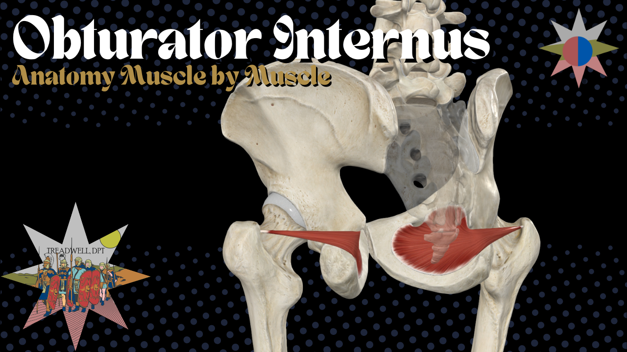
In this episode of Anatomy Breakdown – Muscle by Muscle, we cover:
Obturator internus origin and insertion
Its unique pulley-like path through the pelvis
Innervation and nerve roots
Primary actions
Its role in deep hip stability
Its connection to the pelvic floor
Its relationship with the pudendal nerve and Alcock canal
Clinical considerations for hip and pelvic health rehabilitation
What Is the Obturator Internus?
The obturator internus is one of the deep six external rotators of the hip.
The deep six include:
Piriformis
Superior gemellus
Obturator internus
Inferior gemellus
Obturator externus
Quadratus femoris
These muscles help rotate the femur and provide deep stabilization around the hip joint.
The obturator internus is especially interesting because much of its muscle belly is located inside the pelvis. Its tendon exits the pelvis and bends around the back of the ischium before reaching the femur.
This creates a pulley-like arrangement that redirects the muscle’s line of pull.
Obturator Internus Origin
The obturator internus originates from the:
Internal surface of the obturator membrane
Bony margins surrounding the obturator foramen
Internal pelvic surfaces around the obturator region
The obturator foramen is the large opening in the pelvis located below the acetabulum.
The broad muscle belly of the obturator internus covers part of the lateral pelvic wall before its fibers converge into a tendon.
Obturator Internus Insertion
The obturator internus inserts onto the:
Medial surface of the greater trochanter
Region near the trochanteric fossa of the femur
Before reaching its insertion, the tendon:
Begins inside the pelvis.
Exits through the lesser sciatic foramen.
Bends around the lesser sciatic notch of the ischium.
Travels laterally toward the proximal femur.
Inserts onto the medial greater trochanter.
In simple terms, the obturator internus travels from the internal pelvis to the greater trochanter, with a sharp directional change around the ischium.
Obturator Internus and the Lesser Sciatic Foramen
The obturator internus tendon exits the pelvis through the lesser sciatic foramen.
This is an important distinction from the piriformis:
The piriformis passes through the greater sciatic foramen.
The obturator internus passes through the lesser sciatic foramen.
After exiting the pelvis, the obturator internus tendon changes direction around the ischium. The bony surface acts like a pulley, redirecting the tendon toward its insertion on the femur.
Obturator Internus Innervation
The obturator internus is innervated by the:
Nerve to obturator internus
The associated nerve roots are:
L5, S1, and S2
The nerve to obturator internus also commonly supplies the superior gemellus.
Despite its name, the obturator internus is not innervated by the obturator nerve. The obturator nerve supplies the obturator externus.
Obturator Internus Function
The obturator internus is commonly described as a:
Hip external rotator
When the hip is extended, it helps rotate the femur outward.
Its function changes as the position of the hip changes.
With the Hip Extended
The obturator internus may assist with:
Hip external rotation
Femoral head stabilization
Posterior hip stability
Rotational control
With the Hip Flexed
As the hip moves into flexion, it may contribute more to:
Hip abduction
Deep hip control
Femoral head stabilization
Management of hip rotation
The obturator internus should therefore be viewed as more than a muscle that turns the leg outward. It is part of the deep stabilization system that controls the relationship between the pelvis and femur.
Obturator Internus and Hip Stability
The obturator internus is not a large power-producing muscle like the gluteus maximus.
Instead, it acts more like a fine-control muscle.
It may help:
Center and control the femoral head
Organize hip rotation
Support posterior hip stability
Coordinate movement between the pelvis and femur
Stabilize the hip during weight-bearing activity
This makes it relevant during movements such as:
Walking
Running
Squatting
Lunging
Climbing stairs
Getting into and out of a car
Cutting and pivoting
Single-leg balance
Research using fine-wire electromyography suggests that the obturator internus is active during hip extension, external rotation, and abduction. It may also activate early when the hip needs rotational or frontal-plane control.
Obturator Internus and the Pelvic Floor
One of the most clinically important features of the obturator internus is its relationship with the pelvic floor.
The obturator internus forms part of the lateral wall of the pelvis. The pelvic floor sits below it and helps support the pelvic organs, manage pressure, and contribute to urinary, bowel, and sexual function.
The levator ani has an important fascial relationship with the obturator internus through the obturator fascia.
This means the obturator internus and pelvic floor are not simply located near each other. They are mechanically connected through connective tissue.
Obturator Fascia and Levator Ani
The obturator fascia covers the internal surface of the obturator internus.
Part of the levator ani attaches along a thickened portion of this fascia called the:
Tendinous arch of the levator ani
Arcus tendineus levator ani
Anatomical research has identified broad contact between the obturator internus and levator ani through this fascial region.
This connection suggests that hip mechanics and pelvic floor mechanics may influence one another.
The obturator internus may provide a foundation for part of the pelvic floor, while pelvic floor activity may affect tension and control along the lateral pelvic wall.
Can Hip Strength Affect Pelvic Floor Function?
Research has explored whether strengthening the hip external rotators can influence pelvic floor performance.
Exercise programs involving movements such as clamshells, resisted external rotation, and monster walks have produced improvements in both hip external rotation strength and pelvic floor squeeze pressure in some participants.
This does not mean that hip exercises directly isolate the obturator internus or replace pelvic floor rehabilitation.
It does support a broader clinical concept:
The hip and pelvic floor should not always be treated as completely separate systems.
Hip strengthening may be useful when someone presents with:
Weak hip external rotators
Weak hip abductors
Poor single-leg control
Reduced pelvic stability
Difficulty coordinating the pelvic floor
Urinary symptoms
Pelvic pain
Obturator Internus and Urinary Symptoms
Some research has found weaker hip external rotators and abductors in individuals reporting urinary urgency and frequency.
Interestingly, pelvic floor strength and endurance were not necessarily different from those of participants without symptoms.
This suggests that urinary symptoms may not always be explained by pelvic floor weakness alone.
A complete assessment may also consider:
Hip external rotation strength
Hip abduction strength
Pelvic stability
Single-leg control
Pelvic floor relaxation and coordination
Breathing and pressure management
Bladder habits
Symptom triggers
Obturator Internus and Pelvic Pain
The obturator internus may be involved in some presentations of deep pelvic, hip, or gluteal pain.
Possible symptoms may include:
Deep pelvic aching
Deep buttock pain
Pain near the lateral pelvic wall
Pain with sitting
Pain during hip rotation
Pain during sexual activity
Symptoms that are difficult to localize
Because the muscle is located deep inside the pelvis, discomfort may not feel like a typical superficial muscle strain.
However, these symptoms are not specific to the obturator internus. Pelvic pain can have several muscular, neurological, orthopedic, and medical causes.
Obturator Internus and the Pudendal Nerve
The obturator internus is closely related to the:
Pudendal nerve
Pudendal canal
Alcock canal
The pudendal nerve supplies important sensory and motor function to the pelvis and perineum.
Pudendal nerve irritation may contribute to symptoms such as:
Burning or aching perineal pain
Pain that worsens with sitting
Genital or rectal discomfort
Sexual dysfunction
Urinary symptoms
Bowel symptoms
Altered sensation within the pudendal nerve distribution
The pudendal canal is formed within the obturator fascia covering the obturator internus.
Because of this close anatomical relationship, tension or irritation around the obturator internus may be relevant in some pudendal nerve presentations.
The obturator internus does not automatically cause pudendal neuralgia, but it deserves consideration during a complete pelvic health evaluation.
Obturator Internus vs. Piriformis
The obturator internus and piriformis are both deep hip external rotators, but their anatomy is different.
Piriformis
Originates from the anterior sacrum
Exits through the greater sciatic foramen
Inserts onto the superior greater trochanter
Closely associated with the sciatic nerve
Obturator Internus
Originates around the internal obturator membrane
Exits through the lesser sciatic foramen
Bends around the ischium
Inserts onto the medial greater trochanter
Closely associated with the pelvic floor and pudendal canal
Both muscles contribute to hip rotation and deep joint control, but their locations create different clinical relationships.
Obturator Internus vs. Obturator Externus
The obturator internus and obturator externus are located on opposite sides of the obturator membrane.
Obturator Internus
Located primarily inside the pelvis
Originates from the internal surface of the obturator membrane
Exits through the lesser sciatic foramen
Innervated by the nerve to obturator internus
Obturator Externus
Located outside the pelvis
Originates from the external obturator membrane and surrounding bone
Passes behind the femoral neck
Innervated by the obturator nerve
Can the Obturator Internus Become Tight?
People may describe the obturator internus as tight when experiencing deep hip or pelvic discomfort.
However, the sensation of tightness may reflect:
Increased muscle activity
Protective guarding
Pain-related tension
Muscle fatigue
Reduced hip mobility
Neural sensitivity
Pelvic floor overactivity
A feeling of tightness does not automatically mean the muscle needs aggressive stretching.
The reason for the tension matters.
Obturator Internus Exercises
The obturator internus is difficult to isolate completely because it works with several other hip external rotators.
Exercises used to train the surrounding system may include:
Clamshells
Resisted hip external rotation
Monster walks
Lateral band walks
Bridges
Step-ups
Split squats
Single-leg balance
Controlled squats
Multidirectional stepping
The goal is generally to improve hip control, pelvic stability, strength, and coordination rather than isolate one small muscle perfectly.
Clinical Takeaway
The obturator internus is far more than a small hip external rotator.
It contributes to:
Hip external rotation
Hip abduction in flexion
Femoral head control
Deep hip stability
Pelvic control
Pelvic floor mechanics
Coordination between the hip and pelvis
Its connection with the obturator fascia creates a direct anatomical relationship with the levator ani and pelvic floor.
Its location near the pudendal canal also makes it relevant when evaluating some cases of pelvic pain, sitting intolerance, urinary symptoms, sexual dysfunction, and pudendal nerve irritation.
The obturator internus should not automatically be blamed for every hip or pelvic symptom. However, when symptoms cross between the hip, deep gluteal region, and pelvic floor, this muscle deserves consideration.
It may not be as famous as the piriformis, but clinically, it may be every bit as interesting.
Frequently Asked Questions
What does the obturator internus do?
The obturator internus externally rotates the hip when the hip is extended. In hip flexion, it may assist with abduction and deep hip stabilization. It also helps control the femoral head and coordinate movement between the pelvis and femur.
Where is the obturator internus located?
The obturator internus is located primarily inside the pelvis along the lateral pelvic wall. Its tendon exits through the lesser sciatic foramen and travels toward the greater trochanter.
What is the origin of the obturator internus?
It originates from the internal surface of the obturator membrane and the bony margins surrounding the obturator foramen.
Where does the obturator internus insert?
It inserts onto the medial surface of the greater trochanter near the trochanteric fossa.
What nerve innervates the obturator internus?
The obturator internus is innervated by the nerve to obturator internus, with contributions from L5, S1, and S2.
Does the obturator nerve innervate the obturator internus?
No. The obturator internus is innervated by the nerve to obturator internus. The obturator nerve supplies the obturator externus.
Which foramen does the obturator internus pass through?
Its tendon exits the pelvis through the lesser sciatic foramen.
Is the obturator internus connected to the pelvic floor?
Yes. The levator ani has a close fascial relationship with the obturator internus through the obturator fascia.
Can the obturator internus contribute to pelvic pain?
It may contribute to some pelvic pain presentations because of its location along the pelvic wall and its relationship with the pelvic floor and pudendal nerve. Pelvic pain is multifactorial and requires a complete evaluation.
What is Alcock canal?
Alcock canal, also called the pudendal canal, is a fascial tunnel along the lateral pelvic wall that contains the pudendal nerve and internal pudendal vessels.
Continue Learning Anatomy
Continue exploring the origins, insertions, innervation, actions, and clinical relevance of the deep hip muscles through Anatomy Breakdown – Muscle by Muscle.
Piriformis Anatomy
Gluteus Minimus Anatomy
Gluteus Medius Anatomy
Subscribe to Treadwell, DPT

Fabric: 100% cotton
Fabric Weight:10.3 oz/yd² (350 g/m²)
Fabric Thickness: Thick
For the rehab pros with championship-level hands.
The Soft Tissue Issues Tee brings pro wrestling energy to the physical therapy world with a loud, vintage-inspired graphic built for PTs, PTAs, massage therapists, athletic trainers, students, lifters, and anyone who knows soft tissue work can feel like a main event.
With a bold front logo and a full back graphic featuring championship rehab attitude, this shirt is made for people who live somewhere between the clinic, the gym, and ringside chaos.
Wear it to the clinic, the gym, class, or anywhere you want your PT humor to hit a little harder than the average anatomy tee.
Best for:
Physical therapists, PT students, rehab pros, massage therapists, athletic trainers, gym lovers, wrestling fans, and anyone with soft tissue issues.
NO REFUNDS/EXCHANGES
